


|
Who Was Fortune?
Why Wasn't Fortune Buried?
How Did the Skeleton Get to the Museum?
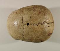
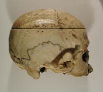
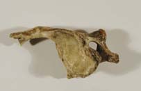
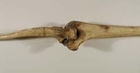
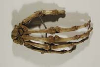
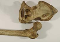
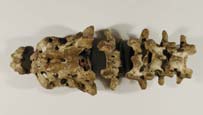
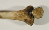
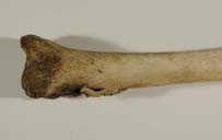
Learning From the Skeleton
The Future For Fortune
Fortune's Story at the Mattatuck Museum
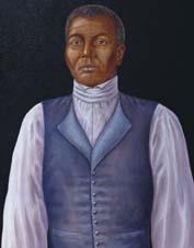
Fortune
Fortune as he may have looked in life. Painted
by William Westwood, a medical illustrator, based on Fortune's skeleton. |
|
||||